THE INTEREST AREAS OF PATHOLOGY

Pathology is generally seen as equivalent to "cancer diagnosis" in the society in parallel with the increasing frequency of cancer. Pathology plays an important role especially in cancer and pathology is the "gold standard" for cancer diagnosis. Many information such as the existence of cancer, its type (differentiation of adenocarcinoma or squamous cell carcinoma with different treatment options in lung cancer, differentiation of lymphoma and carcinoma in gastric cancer, cervical / endometrial cancer distinction in uterine cancer, etc.), classification, parameters required in treatment and follow-up, treatment response are determined by pathological examination. The most important step in cancer treatment is the accuracy of pathological diagnosis.

  • Non-cancer diseases: Gastroenterological diseases, endoscopic / colonoscopic examinations, diagnosis of dermatological diseases, bone marrow and lymph node examinations for the diagnosis of hematological diseases, diagnosis of gynecological and rheumatological diseases, liver and kidney diseases, all kinds of tissue samples taken from the body can be pathologically examined and diagnosed.
  • Follow-up biopsies: Pathological examination is required for the diagnosis of biopsy samples of patients undergoing organ transplantation, various conditions that occur after transplantation and complications.
  • Cancer screening: In cervical cancer, pathological evaluation as in PAP test / PAP smear examination is performed to diagnose and treat diseases before cancer development. It is determined how the patient will be followed and treated according to the PAP smear result.
  • Non-surgical biopsies:Nowadays, with the development of interventional radiological techniques, samples can be taken from almost all organs by radiology-supported cytological examination and tru cut biopsies can be performed. In such procedures, pathology both evaluates the adequacy of the biopsy at the bedside and determines the diagnosis of the disease with the next stages.
  • Pathological examination in the operating room environment: Urgent pathological examination is also possible during the operation. In the presence of unexpected findings or to determine the framework of the surgery, while the surgical procedure continues, pathologists determine the course of the surgery by evaluating.

Pathology examines 4 main themes related to a disease and all the necessity of evaluating these stages in order to recognize them correctly.

Etiology

Cause: The causative agent

Pathogenesis

Occurrence: The path followed by the agent in creating the disease

Morphology

Appearance: Structural changes that occur as a result of the disease

Clinic

Functional change caused by the structural change in the patient

STEPS OF THE PATHOLOGICAL EXAMINATION

The final product of the pathology is the "Pathology Report". There is a lot of human effort and multiple laboratory steps behind this pathology report, which is often traced in a single page document. The indispensable part of pathology is human and human labor. Memorial Pathology; carries the journey of pathology starting with microscope to artificial intelligence and robotic applications and crowns human labor with its technological infrastructure. Memorial pathology laboratory has digital workflow and each step is followed digitally in the laboratory.

Idendification

Identification is essential at all stages in pathology. In Memorial Pathology, every stage of the laboratory is followed by a QR code and it is ensured that the error is minimized by digitally monitoring which work is done in each step.

Fixation

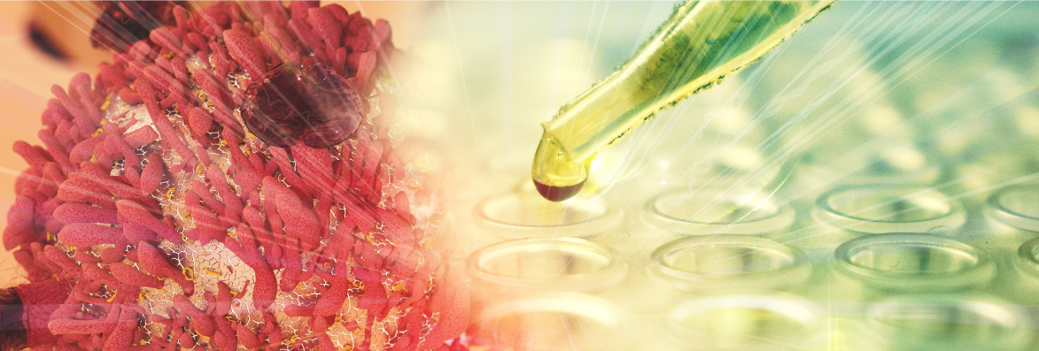
In order to prevent the tissues from losing their vitality and becoming unrecognizable, some procedures are applied during tissue processing. First of all, macroscopic examination is performed on the tissues fixed in a liquid called "formaldehyde".

Macroscopic examination

In pathology, the surgical material is examined manually and visually. The macroscopic examination stage allows 50% of the diagnosis to be made.

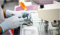
Tissue processing and preparation of paraffin blocks

The sampled tissues are then taken to the "tissue processing’ and during this 12-hour process, tissues are passed through chemicals such as acetone and alcohol and the water inside the cell is taken and paraffin is ensured to penetrate into the tissue.

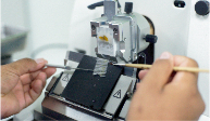
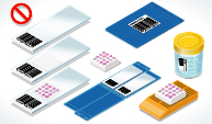
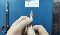
Taking sections from paraffin blocks

Sections of 3-4 micrometer thickness are taken from the paraffin blocks with a microtome and these are stained to be visible after they are placed on a 'glass slide'. The dye used in pathology routine is 'Hematoxylin-Eosin' dye.

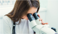
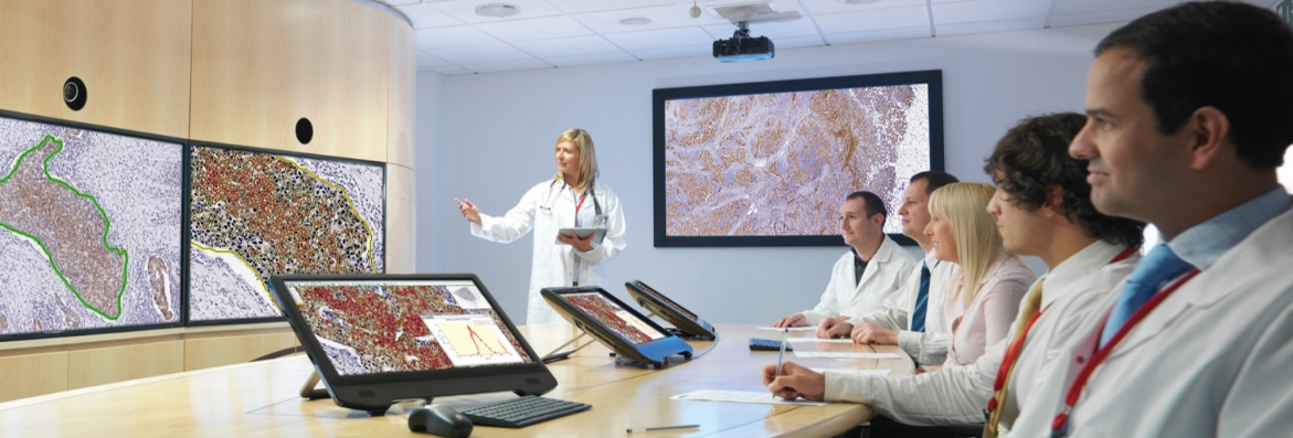
Microscopic examination

The glass slides that have been completed the laboratory stage are ready for microscopic examination and the morphological changes in the cells are evaluated under the microscope by the pathologist and the disease is diagnosed.

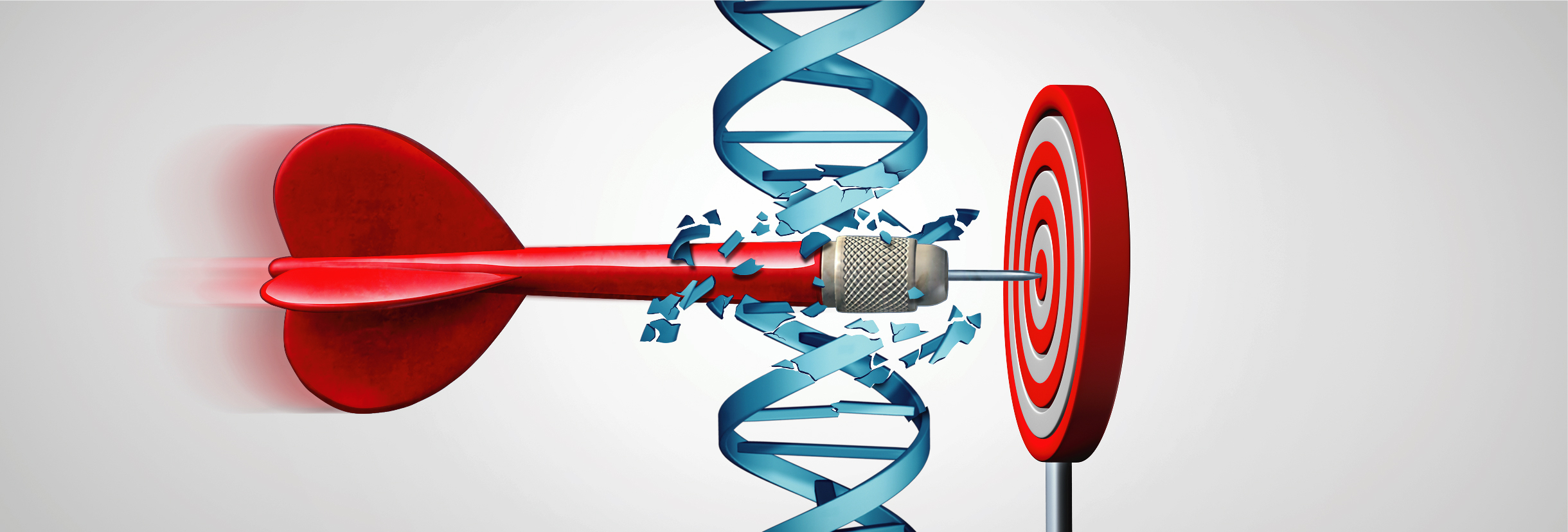
Additional examinations

After the initial evaluation of routine Hematoxylin-Eosine stained sections, additional examinations may be required according to the preliminary diagnoses. The aim here is to enable further investigations by revealing the origins and properties of cells.

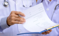
Preparation of Pathology Report

Pathology report includes identity information, report information, macroscopic findings, microscopic findings, pathological diagnosis, specific examinations, epicrisis / explanation, intraoperative consultation result.
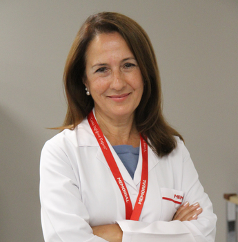
Our Doctors

Together with our consultant physicians

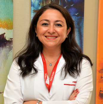
Prof. İlknur TÜRKMEN

Gynecopathology, Digital Pathology, Dermatopathology
Prof. Fatma Aktepe

Breast Pathology
Uropathology
Prof. Gülen Bülbül Doğusoy
Digestive System/Liver and Pancreas - Head/Neck and Cardiovascular Pathology
Prof. Türkan Atasever Rezanko
Cytopathology, Neuropathology, Bone soft tissue and Head/neck Pathologyi
Assoc. Prof. Pembe Gül
Güneş
Dermatopathology, Endocrine system pathology, Gynecopathology
Prof. Şemsi YILDIZ

Hematopathology, Gynecology,
Gastrointestinal tract pathology
Doç. Dr. Serdar Balcı (K)

Liver/pancreas and Digestive system pathology, Uropathology, Neuropathology
Doç. Dr. Murat Oktay


Surgical pathology

Prof. Mehtat Uz Ünlü (K)
Nephropathology, Gastrointestinal System Pathology, Head and Neck Pathology and Thyroid Pathology, Laboratory Accreditation
Prof. Sülen SARIOĞLU

Nephropathology, Head and Neck Pathology
Prof. Emine Çağnur ULUKUŞ

Gynecopathology, Lung Pathology

Exp. Dr. Emre Karakök

Surgical pathology

Exp. Dr. Fadime Gül SALMAN

Gynecopathology,
General Surgery Pathology
Exp. Dr. Fatma Gülgün Sade Koçak (K)
Surgical pathology, Gastrointestinal System Pathology, Gynecology
Exp. Dr. Rukiye Nilgün Erdoğan

GIS Pathology, Lung Pathology, Cytopathology
Exp. Dr. Nural ÖREN
Gastrointestinal System Pathology

FREQUENTLY ASKED QUESTIONS

The pathology report contains a lot of information necessary to organize the treatment of a patient with cancer. Frequently, treatment protocols are determined in the light of the information provided in the pathology report. The treatment of the disease is arranged according to clinical course and various parameters such as the "stage of the tumor", mode of behavior, its degree of spread in the body. Pathological examinations are also required to determine a number of treatment options, called "targeted therapy", which reduce the side effects of chemotherapy.
Cancer cells try to resemble the tissues they originated from. This similarity is expressed as "differentiation" and is defined by grade. Low-grade tumors are well differentiated, that is, more similar to the tissue they originated from, while high-grade tumors are less differentiated and their resemblance to the tissue from which they originate may sometimes be scarcely any. The stage of cancer is a term that defines its spread in the body. While stage 1 tumors are limited in the organ they originated from, the presence in the body increases as the stage increases.
Molecular pathology techniques are used to determine suitability for targeted therapy. Some drugs are used in the treatment in the presence or absence of related mutations. While these analyzes determine the patients who may benefit from the drug, they also identify the patients that will not be benefited from the drug and protect these patients from the possible side effects of the drugs.
Cancer patients are living longer now and new treatment methods have become routine over the years. Therefore, tumor tissue has become more valuable for patients than before. Potentially, molecular pathology studies can be performed on any tumor. However, in this case, care should be taken to preserve the tissue that can be used in the future. Molecular studies should not be conducted if the result to be obtained will not be used in the treatment of the patient (ie there is no clinical indication) or if this analysis is not part of a scientific study approved by the ethics committee.
Tissues taken by surgery and biopsy are not completely composed of tumors. They may also include areas of normal tissue, necrotic dead tissue, or tumor precursor (such as adenoma, in situ tumor). Subgroups are also formed within the tumor, and while some subgroups are the main cause of the disease, it grows rapidly, while others may have lost their ability to grow. A competent pathologist in tumor pathology should select the appropriate area of the tumor by microscope and indicate the number or percentage of tumor cells in the chosen area. In cases where this preliminary evaluation is not made, false positive or false negative results are obtained, patients who need to receive treatment cannot receive the treatments, and patients who are not expected to benefit from treatment are exposed to the drug unnecessarily.
Molecular studies can be performed on paraffin-embedded tissues (wax blocks) and tissues stored with rapid freezing (-80 C) with appropriate detection and tracking. For this reason, proper determination of tissues taken by surgery or biopsy (taking them into formaldehyde solution very quickly) is very important. Molecular studies will not be healthy, as there will be autolysis in tissues that are not detected with sufficient formaldehyde.
If appropriate detection and follow-up procedures are applied, molecular pathology techniques can be performed even after decades from tissues stored in appropriate pathology archives.
Molecular pathology reports usually contain detailed technical information. The presence or absence of a mutation may indicate the possibility of responding to treatment in some diseases, while it may indicate that treatment should not be given in some diseases. Therefore, molecular pathology reports should be interpreted by the oncology specialist who follows the patient.
Although they are generally used for tumors in molecular pathology, similar techniques can be applied for infectious diseases and identification.
Different techniques have different specificity and sensitivity in molecular studies. For this reason, tests specific to the patient and the planned treatment should be selected.
For molecular techniques, determination and follow-up procedures suitable for tissues should be performed and stored in appropriate archives in pathology laboratories. If you are transporting paraffin blocks (wax blocks) to another lab, you should keep them at room temperature (21-23 ° C).
For cancer patients, multidisciplinary meetings attended by all doctors who have a role in the diagnosis and treatment of the patient are called the "Tumor Council". Pathology, the gold standard of cancer diagnosis, is also a permanent member of tumor councils. In addition to diagnosis, pathology has a role in many points such as prognostic parameters, treatment options, treatment responses, and the decision of additional examinations to be performed for the patient. Our pathologists for their care services.

CONTACT US