Pathology is a clinical branch that includes laboratory service. It provides a kind of 'consultation service' to other clinical branches and enables the diagnosis of diseases. Therefore, when examining under the microscope, the pathologist must know not only the image, but also the clinical condition of the patient, when the disease started, and what diseases the person had before.
How does the pathology laboratory work?
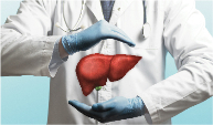
Samples required for pathological examination must undergo an effective and detailed laboratory study in order to reach the process of working with a microscope. Specific pathology services include both anatomical (surgical pathology, cytopathology, autopsy examination) and clinical pathology (nephropathology, hepatopathology, etc.).
The pathological examination, which requires a long kitchen work in the pathology laboratory, emerges as a 'pathology report' after the pathology specialist doctor's evaluations under the microscope.
A very meticulous study is required in the laboratory part of the pathology, and this stage directly affects the diagnosis of the disease.
In order for the pathology report to reach the patient, it is essential that the pathologist evaluates it at the microscope. However, before this stage, there is a laboratory and preparation stage carried out by pathology technicians, which requires a lot of effort. This stage constitutes the kitchen of pathology and requires pathology specialists and pathology technicians to work in harmony. Pathology technicians are one of the keystones of the pathology laboratory.
A very meticulous study is required in the laboratory part of the pathology, and this stage directly affects the diagnosis of the disease. Correct diagnosis in pathology practice; It depends on the standardization of the laboratory stages and the correct execution of all steps.
Authentication
Authentication is essential at all stages in pathology. Every stage of the laboratory at Memorial Pathology
It is followed with a QR code and the work done at each step is digitally monitored, minimizing the error.
Fixation
Some procedures during tissue detection so that the tissues do not lose their vitality and become unrecognizable.
is implemented. First of all, macroscopic examination is performed on the tissues fixed in a liquid called 'formaldehyde'.
Macroscopic Examination
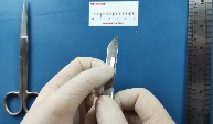
In pathology, the surgical material is examined manually and visually. Macroscopic examination phase, diagnosis
It allows 50% to be placed. Then, appropriate samples that will show the disease and lead to the diagnosis are taken from the relevant organ. If a sample is not taken from the right place, it is not possible to make a correct diagnosis.
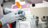
Tissue Tracking And Preparation Of Paraffin Blocks
The sampled tissues are then taken to the “follow-up” process and
In the process, the water in the cell is taken and passed through chemicals such as acetone and alcohol, allowing paraffin to penetrate into the tissue. In other words, a kind of tissue is mummified and made 'immortal'. Tissues turned into “paraffin blocks” become a material that can be examined after 20 years.
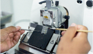
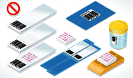
Taking Sections From Paraffin Blocks
Sections are taken from paraffin blocks with a microtome device when they are 3-4 micrometers thick, and these are
Once placed on the 'slide' it is painted to make it visible. The dye used in the pathology routine is the 'Hematoxylin-Eosin' dye.

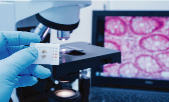
Microscopic Examination
After the laboratory phase is completed, the slides are ready for microscopic
examination and are placed under the microscope. The morphological changes in the cells are evaluated by the pathologist and the disease is diagnosed.

Additional Reviews
After the initial evaluation of routine Hematoxylin Eosin stained sections, additional investigations may be required in line with the preliminary diagnosis.
The aim here is to provide the origins and characteristics of the cells for further examination
These examinations can be Histochemical Examination, Immunohistochemical Examination, Flow Cytometry, (in)direct Immunofluorescence Examination, In Situ Hybridization. For Detailed Information About Additional Reviews
Pathology materials have the ability to be stored and re-examined when desired.
For example; When a new treatment method emerges after diagnosis for a patient undergoing cancer treatment, a new treatment opportunity can be provided for the patient by performing molecular pathological examinations from the previous tissue.
For Detailed Information About Molecular Pathology
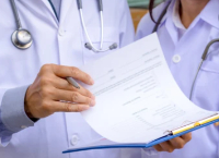
Preparation of Pathology Report
Pathology report; identification information, report information, macroscopic findings, microscopic findings,
pathological diagnosis,
Includes special investigations, epicrisis/explanation, intraoperative consultation result.
For detailed information
Second Opinion/Consultation in Pathology
Pathology serves all clinical branches. With such a wide area of interest and such a shortened half-life of information, branching in pathology has become indispensable. Memorial Pathology produces reports at international standards with its physician staff specialized in their fields.
For Physician Staff Information
In order to get a second opinion in pathology, it is necessary to examine the slides and/or paraffin blocks of the biopsy materials. The patient/physician can always ask for a second opinion in line with medical necessity. Paraffin blocks are stored for 20 years.