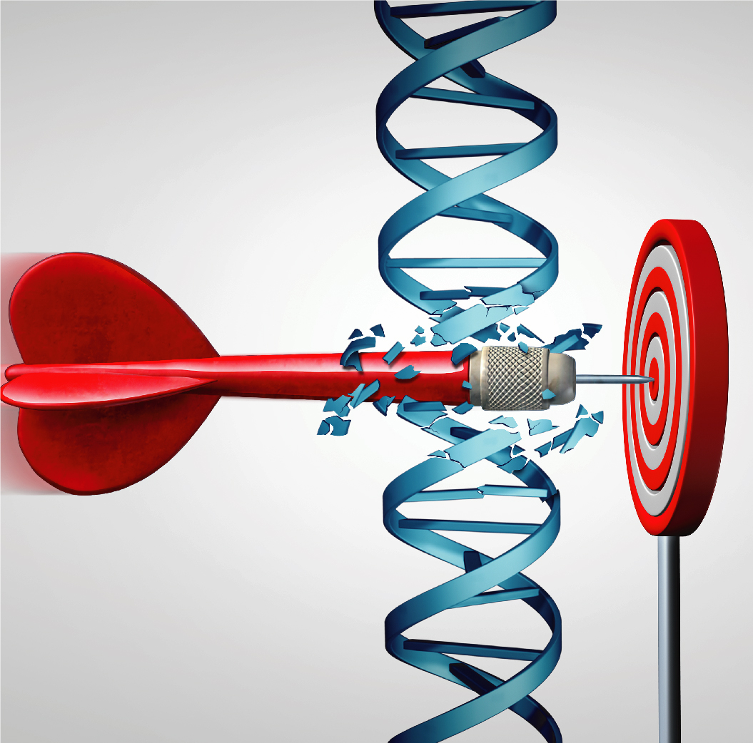
Uses of molecular techniques
Molecular techniques are used for different purposes in pathology.
It is used to diagnose tumors or infectious diseases with appropriate tests after the morphological evaluation of a competent pathologist and to determine tumor subtypes. Complementary morphology and molecular examinations have gradually become an integral part of each other in the diagnosis/follow-up and treatment of oncological patients. It is in such a manner that, the names of some tumors (brain tumors and various sarcomas) were changed by comparing molecular techniques and morphologies, and new diagnostic groups were created.
The use of targeted therapies is possible with the presence or absence of mutation in cancerous tissues. Detection of the presence or absence of these mutations is made with tests applied in molecular pathology. For example; EGFR, ALK, ROS1 tests in lung cancer, KRAS, NRAS, BRAF tests in colon cancer, Her2 test in breast cancer and stomach cancer, BRAF test in thyroid cancer and skin cancer are among the routine tests of oncology.
Although the course of the disease is very dependent on individual factors, it is necessary to know the possible course of the disease for appropriate treatment management for the patient. Therefore, some molecular techniques used guide oncologists. For example; MSI (Microsatellite Instability) tests, which show the possibility of hereditary cancer in uterine cancer, are now routinely applied in pathological examination.
Techniques Used in Molecular Pathology
Similar to the immunohistochemical studies used in Daily pathology practice, it allows the determination of protein structures that occur with mutation. Because it is fast and practical (such as BRAF in thyroid and skin cancer, IDH1 in brain tumors), it is used in the detection of some mutations.
They are tests performed using special opposite (complementary) DNA and RNAs that bind to DNA or RNAs in cells. It is used in etection of viruses (EBER in various lymphomas, HPV in cervical cancer), detection of tumor focus (albumin in liver), detection of chromosomal changes (1p19q loss in brain tumors, BCR-ABL fusion in some lymphomas) and detection of increase in gene expressions (HER2, EGFR in breast and stomach cancer).
FlSH (Fluorescent in-situ hybridization):
These are in-situ hybridization examinations performed using special fluorescent luminescent kits and special fluorescent microscopy.
CISH (Chromogen in-situ hybridization): These are the examinations that enable in-situ hybridization to be performed with light microscopy.
Polymerase chain reaction (Polymerase Chain Reaction) is the process of reproducing DNA or RNAs obtained from tissue in the laboratory. If there are mutations in a certain number of replication times, sufficient new DNA or RNA is formed and the presence of mutations (for example, KRAS, NRAS, BRAF tests in colon cancer / bowel cancer) are detected.
The mutations seen in tumors are very diverse. While some mutations are the main factor in the proliferation of the tumor, some remain only with change and do not affect the behavior (tumor biology) of the tumor. These "driver" mutations that affect tumor behavior and "passenger" mutations that do not affect the behavior are different in terms of diagnosis, treatment and survival. For this reason, analyzes specific to selected mutations (point mutation analyzes) that can directly determine tumor behavior are used in molecular pathology.
It is an analysis made by reading the bases in a DNA sequence one by one. Thus, it is understood with which change the mutation occurred and the diagnosis and treatment selection is made accordingly.
All mutations in a tumor can be detected by new generation sequencing based on reading multiple foci of DNA at the same time. It is used to detect mutations that are not frequently seen in the relevant tumor, but have medications for treatment, especially in patients with advanced stages and who have completed their first treatments. The system, which is located in a limited number of centers in Turkey, is used in the ISO 15189 accredited Memorial Genetics Laboratory.
Changes in DNA are called mutations. Mutations occur in almost every cell division and almost all of them are repaired. These mutations accumulate because cancer cells grow too fast and their repair mechanisms cannot make adequate corrections. The mutation burden measurement obtained by the ratio of this mutated accumulation to the total amount of DNA has recently been used in treatment selection. The more mutations there are in a tumor (such as skin cancer / melanomas), the more foreign antigens will be produced and these tumors will respond better to immunomodulatory treatments (PD1-PDL1 tests used in many types of cancer, especially lung cancer).